A Doctor From ITS Develops 3D Imaging System for Bone Contour
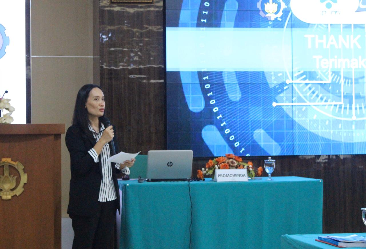
Tita Karlita when presenting her dissertation at the Open Session of Doctoral Promotion in the Department of Electrical Engineering ITS.
ITS Campus, ITS News – In helping to develop the imaging function in the medical world using ultrasound, a student of the doctoral program of the Institut Teknologi Sepuluh Nopember (ITS) designed an ultrasound imaging system to reconstruct bone outer counters in 3D. This topic was raised in a dissertation presentation at the Open Session of Doctoral Promotion in the Department of Electrical Engineering ITS, last Tuesday (25/2).
Tita Karlita explained, usually computerized tomography (CT) was recognized as the good standard in bone imaging. However, while using X-ray, explained Tita, this method has a high radiation exposure. “Because of that, the frequency of use for humans is limited,” she said.
Based on Tita’s explanation, nowadays ultrasound is not recommended for bone imaging yet. However, she explained that ultrasound has the advantage of not emitting radiation, available in various medical centers, and cheaper. Based on those reasons she determined to take this research topic to get her doctorate.
Through her dissertation entitled Reconstruction of Long Bones Using 3D Ultrasound Imaging System, Tita introduced NEURON, a 3D imaging system using ultrasound. The lecturer from Informatics Department at Politeknik Elektronika Negeri Surabaya (PENS) explained that NEURON is a combination of two methods: the Regent Proposal Network (RPN) and the Curve Approximation.
Tita explained her research which was guided by Prof. Dr. Ir Mauridhi Hery Purnomo MEng, Dr. I Ketut Eddy Purnama ST MT, and Dr. Eko Mulyanto Yuniarno ST MT, that using RPN is one of the deep learning methods. This method serves to minimize the bone detection area. Continued Tita, the results were extracted using the Curve Approximation method. “The results of the merger of the two obtained by 3D imaging of the contours outside the bone,” she said.
Related to the difference with 3D Ultrasound which is widely used in hospitals, Tita said that its behind the scenes work is almost the same. The difference is that 3D Ultrasound only does the scanning and reconstruction without segmentation. In the other side, NEURON tries to eliminate disorders such as muscles or tendons so that the purpose of bone contour reconstruction can be accomplished. “It’s different with the 3D Ultrasound which aims to scan organs,” she said.
An example application of 3D bone contour imaging is in the field of forensic anthropology. Tita explained, anthropology method uses bone as one of the objects to identify individuals. The field requires a lot of data. “Because CT Scan has limitations in taking a lot of samples, ultrasound can be an alternative,” she concluded. (fat/sel/ITS Public Relation)
Related News
-
Supporting the Development of Material Processes, ITS Professors Utilize Biomass Waste
ITS Campus, ITS News — Innovation in material processes continues to develop to support society’s need for environmentally friendly
July 10, 2020 14:07 -
ITS Professor Ideas for Bio-Corrosion Control in Marine Structures
ITS Campus, ITS News — Coastal and offshore buildings interacting directly with seawater trigger damage, including bio-corrosion. If not
July 10, 2020 14:07 -
ITS Launches the First Marine Floating Solar Power Plant Prototype in Indonesia
ITS Campus, ITS News — Institut Teknologi Sepuluh Nopember (ITS)‘s commitment in realizing the energy transition is getting serious.
July 10, 2020 14:07 -
Targeting Champion, Bayucaraka ITS Launches Flying Robot Innovation
ITS Campus, ITS News – The Bayucaraka team at Institut Teknologi Sepuluh Nopember (ITS) has launched four new flying
July 10, 2020 14:07